Endoplasmic Reticulum and Golgi Apparatus: The Added Value on Protein Synthesis
Episode #5 of the course How the cells work by Luis Francisco Cordero
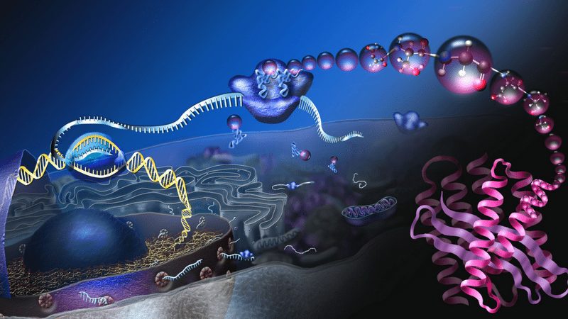
Proteins are more, much more than strings of amino acids. Protein synthesis is fascinating, but the incorporation of many amino acids in the precise order dictated by our DNA is not nearly enough for them to perform the amazing tasks they are in charge of. They must be turned, twisted, polished in many ways, and sometimes even attached to other proteins to function properly.
Just a Twist and a Turn?
It is far more complicated than that yet simpler in some way. While there are molecular compounds that aid in the process, for the most part, protein folding into the specific 3-dimensional shape required for them to work is self-directed. That is, proteins perform this elaborate act of acquiring a specific shape mainly by themselves. How? Due to the positive and negative charges in their amino acids, they attract and repel other parts of the chain. Amazing as it can be, some portions of the amino acids can interact with parts of others, and this results in a very predictable folding pattern.
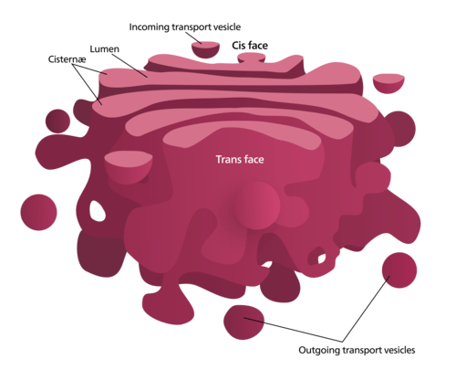
Golgi apparatus
At the same time, it is still more complicated than just that. Many proteins have to be cleaved and re-assembled, and some of them need to attach sugar or fat molecules in order to become fully functional. For many, this complex post-processing occurs in a fascinating network of tubular structures that are called the endoplasmic reticulum and Golgi apparatus. Here, many proteins complete their maturation when some of their amino acids get fastened with other compounds.
Apart from being highly specialized post-production cellular facilities, these organelles oversee newly synthesized protein sorting. That is, they “decide” which proteins stay on those compartments and which ones migrate to another organelles, to the plasma membrane, or even to the surrounding milieu. Whenever a molecule is designed to be transported outside the cell, we call this process “secretion.”
Did You Know?
Today, visualization of microscopic subcellular (tinier than a cell and located within its boundaries) has greatly advanced, and membrane-bound organelles such as the Golgi apparatus can be explored and presented in all their glory. But that was not the case when Camillo Golgi discovered it at the turn of the 20th century. His finding was largely criticized, as many fellow scientists of the time considered it an artifact, a false image arising from the limited microscopy techniques that were available back then.
Key Takeaways
Proteins are made according to what our genetic code dictates, but this is not enough for them to work properly. Their full potential is only attained after a kind of post-processing. That is, after the primary chain of amino acids has been assembled, it has to be bent in a very meticulous 3-dimensional shape that is particular to each protein, depending on their function. Also, sometimes it is required that simple carbohydrate or fat molecules be attached to the protein for it to be completely mature and ready for work. This happens in the endoplasmic reticulum and Golgi apparatus. In the latter, many proteins are also sorted and assigned to be distributed to other organelles, the plasma membrane, or even secreted outside the cell.
Tomorrow, we’ll learn about “inner world” of each cell: the cytoplasm.
Recommended book
The Machinery of Life by David S. Goodsell
Share with friends