
How Do We Move?
Episode #8 of the course Understanding your brain by Betsy Herbert
Welcome back!
Until now, we’ve been looking at all the different sorts of incoming signals that the brain interprets as sensation. But what about the outgoing signals? How can we act upon and speak about these things we see? Today, we’re looking at how the brain generates and controls movement.
Take a simple movement, such as climbing the stairs or catching a ball. Seems easy, right? In fact, to carry out even the most basic of movements, the brain has to perform a colossal number of calculations to keep track of your limbs in space, to make sure you don’t miss the ball, close your fingers too fast, or slam your foot down too hard. For all this complex planning and guidance, we have our motor cortex to thank.
The motor cortex is located just behind the somatosensory cortex we saw in Lesson 5. And like the somatosensory cortex, it also has a “body map,” a complete projection of all the areas in our body that contain muscles. When you want to move a certain muscle, a signal is generated in the region of the motor cortex that corresponds to that body area, and is sent down the spinal cord and onto the motor neurons that innervate the muscle itself. The synapse between the tip of the motor neuron and the muscle is rather special: It’s called the neuromuscular junction, and it uses acetylcholine as its neurotransmitter. When the signal reaches this synapse, acetylcholine is released onto the muscle, causing calcium ions to be released, which triggers the muscle to contract. And voilà, movement!
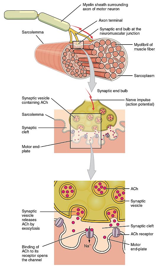
Signal transmission from nerve to muscle at the motor end plate.
Ah, if only it were that simple. In fact, the motor cortex doesn’t work alone in producing these signals—and if it did, all our movements would be jerky, arrhythmic, and uncoordinated. Many other brain regions contribute to modify the signal at different stages on its journey to the spinal cord. The premotor area and supplementary motor area are involved in preparation and planning for movement; the parietal cortex keeps track of where your limbs are in space; the basal ganglia acts as a complex filter, choosing which muscles to inhibit while others contract; and the cerebellum performs all the fine-tuning and learning to allow our movements to become smooth and skilful. You become all too aware of the role of your cerebellum if you drink alcohol; cerebellar function is among the first to be inhibited, meaning your movements quickly become uncoordinated and sloppy.
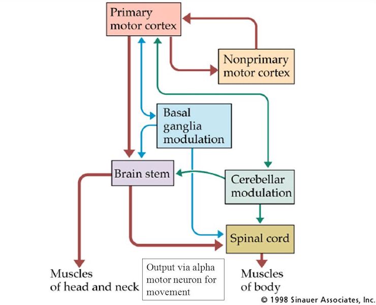
Interactions of CNS areas involved in movement.
By now, you’ll be starting to get an idea of how intricate this system is. But movement isn’t only generated at the level of the brain. For quick and reflexive movements, like dropping a hot object, the signal is generated in the spinal cord itself, stimulated directly via sensory neurons in a loop known as the reflex arc. Rhythmic movements such as walking and running are also largely generated in the spinal cord, via a network of oscillatory neurons known as central pattern generators. And even at the muscle itself, the signal can still be modified: Receptors inside the muscle called muscle spindles detect the degree to which the muscle is being stretched and send feedback signals back to the motor neuron when—ow!—it gets too much.
That’s it for our tour of the motor system today. As you head to the kitchen and make that next cup of coffee, hopefully, you’ll be thinking of all the brilliantly complex processes that are happening in your brain while you do so.
And speaking of coffee, how does it give us that extra buzz? How does any drug affect the brain? Let’s find out tomorrow.
See you then!
Betsy
Recommended reading
Share with friends